Copyright
©The Author(s) 2019.
World J Gastrointest Oncol. Dec 15, 2019; 11(12): 1231-1239
Published online Dec 15, 2019. doi: 10.4251/wjgo.v11.i12.1231
Published online Dec 15, 2019. doi: 10.4251/wjgo.v11.i12.1231
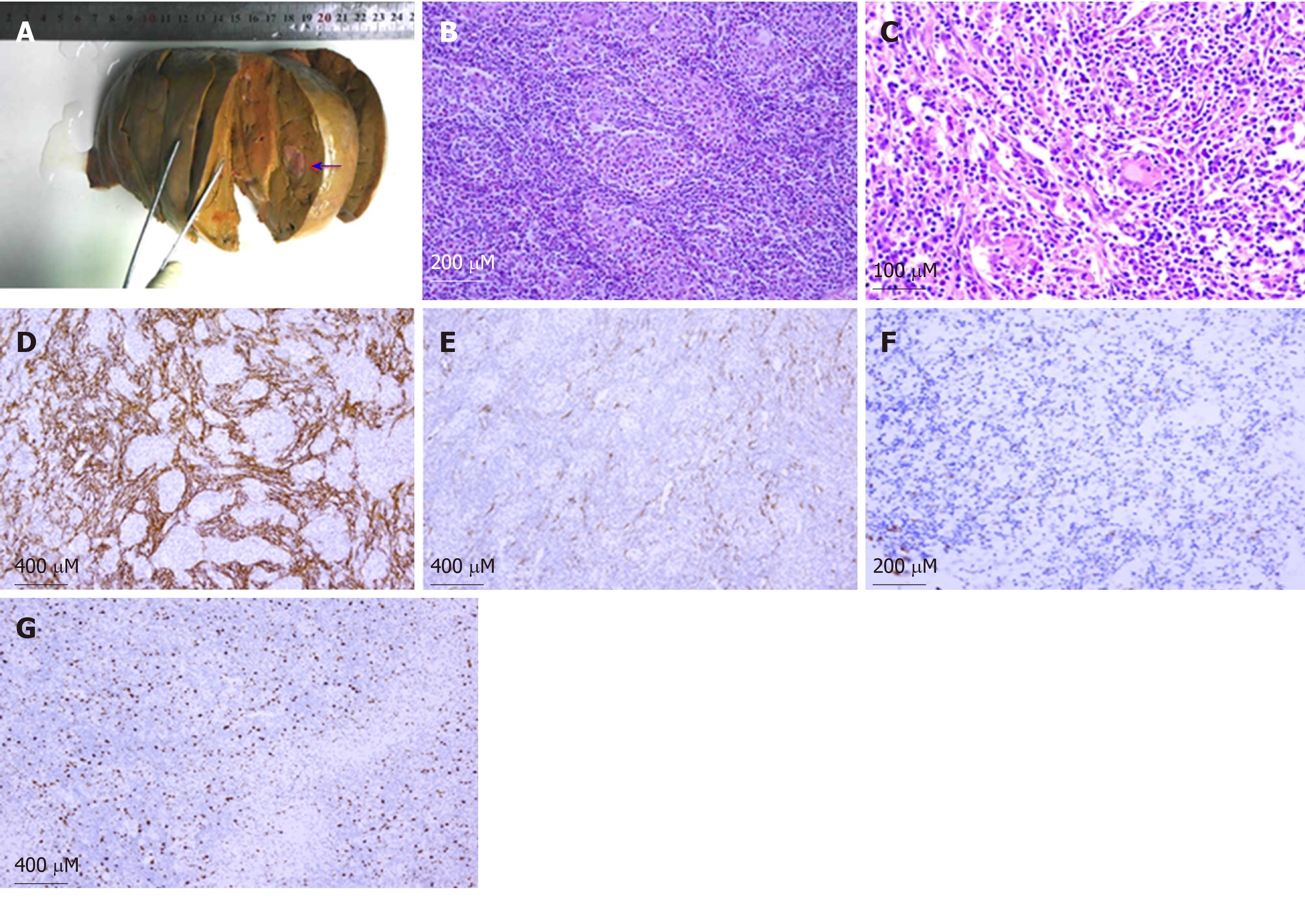
Figure 3 Epstein-Barr virus-positive inflammatory pseudotumor-like follicular dendritic cell sarcoma in the liver.
A: Gross picture of an inflammatory pseudotumor-like follicular dendritic cell sarcoma of the liver. A well-circumscribed solid nodule was found in the liver. Note the grayish-white colored and soft cut surface with focal hemorrhage (arrow); B: Haematoxylin and eosin stained image showing that the tumor tissue had a meshwork-like architecture (× 200); C: On high-power field, the tumor was composed of oval to spindle cells with vesicular chromatin and distinct nucleoli. There was less degree of atypia. The background showed abundant lymphocytes and plasma cells (× 400); D: CD21 was detected on the membrane of almost all of tumor cells by immunohistochemistry (× 100); E: Smooth muscle actin was detected in the cytoplasm of a part of tumor cells by immunohistochemistry (× 100); F: Epstein-Barr virus-encoded small RNA-based in situ hybridization demonstrated positive nuclei of the neoplastic dendritic cells (× 200); G: Ki-67 was detected in the nuclei of almost all of tumor cells by immunohistochemistry (30%; × 100).
- Citation: Zhang BX, Chen ZH, Liu Y, Zeng YJ, Li YC. Inflammatory pseudotumor-like follicular dendritic cell sarcoma: A brief report of two cases. World J Gastrointest Oncol 2019; 11(12): 1231-1239
- URL: https://www.wjgnet.com/1948-5204/full/v11/i12/1231.htm
- DOI: https://dx.doi.org/10.4251/wjgo.v11.i12.1231


