Copyright
©The Author(s) 2018.
World J Gastrointest Oncol. Sep 15, 2018; 10(9): 231-243
Published online Sep 15, 2018. doi: 10.4251/wjgo.v10.i9.231
Published online Sep 15, 2018. doi: 10.4251/wjgo.v10.i9.231
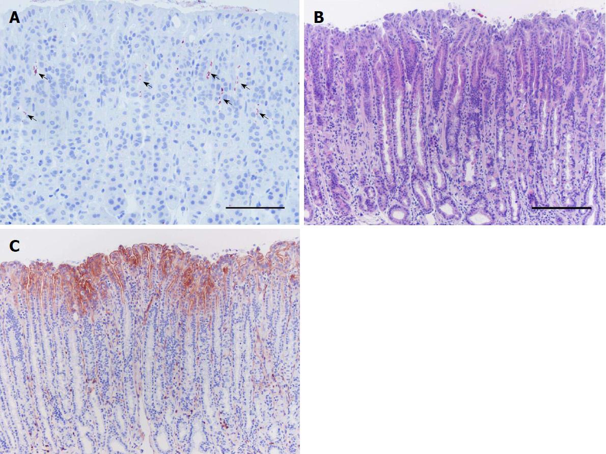
Figure 3 Plasminogen activator receptor induction in gastric epithelial cells in response to Helicobacter pylori infection.
Stomach tissue sections of a mouse infected with H. pylori and euthanized 14 wk after inoculation processed for immunohistochemistry against H. pylori (A) and uPAR (C), and with H&E staining (B). Clusters of H. pylori bacteria (arrows) are observed in the upper third of the gastric glands along the gastric epithelium of the mouse stomach (A). Histopathological alterations are seen, including inflammation and mucous metaplasia (B). uPAR expression becomes evident at the apical membrane of foveolar epithelial cells in the corpus epithelium of H. pylori-colonized mice, such as the representative immunohistochemistry staining shown here (C). uPAR-positive scattered neutrophils are seen in the microphotograph (C) since they constitutively express this molecule. Scale bars: A: 100 μm; B and C: 200 μm. H. pylori: Helicobacter pylori; H&E: Hematoxylin and eosin; uPAR: Plasminogen activator receptor.
- Citation: Molina-Castro S, Ramírez-Mayorga V, Alpízar-Alpízar W. Priming the seed: Helicobacter pylori alters epithelial cell invasiveness in early gastric carcinogenesis. World J Gastrointest Oncol 2018; 10(9): 231-243
- URL: https://www.wjgnet.com/1948-5204/full/v10/i9/231.htm
- DOI: https://dx.doi.org/10.4251/wjgo.v10.i9.231


