Copyright
©The Author(s) 2018.
World J Gastrointest Oncol. Mar 15, 2018; 10(3): 91-95
Published online Mar 15, 2018. doi: 10.4251/wjgo.v10.i3.91
Published online Mar 15, 2018. doi: 10.4251/wjgo.v10.i3.91
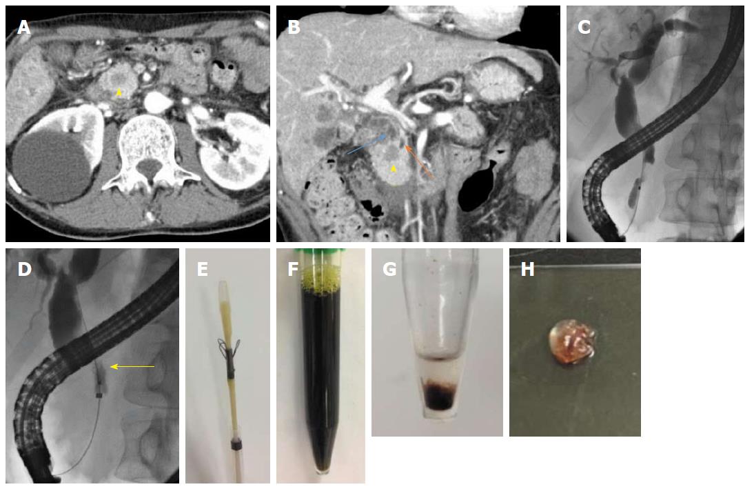
Figure 1 Imaging findings and samples obtained using the Trefle® device.
A and B: Abdominal computed tomography indicated a poorly enhanced region (yellow arrowhead), dilated common bile duct (blue arrow), and upstream main pancreatic duct (orange arrow); C: Endoscopic retrograde cholangiopancreatology demonstrated a biliary stricture; D: The Trefle® device was inserted and opened, and the scraping loops were identified under fluoroscopic guidance (yellow arrow); E: Appearance of the Trefle® device; F: Appearance of samples obtained; G and H: Appearance of the centrifuged deposit.
- Citation: Kato A, Naitoh I, Kato H, Hayashi K, Miyabe K, Yoshida M, Hori Y, Natsume M, Jinno N, Yanagita T, Takiguchi S, Takahashi S, Joh T. Case of pancreatic metastasis from colon cancer in which cell block using the Trefle® endoscopic scraper enables differential diagnosis from pancreatic cancer. World J Gastrointest Oncol 2018; 10(3): 91-95
- URL: https://www.wjgnet.com/1948-5204/full/v10/i3/91.htm
- DOI: https://dx.doi.org/10.4251/wjgo.v10.i3.91


