Copyright
©The Author(s) 2018.
World J Gastrointest Oncol. Dec 15, 2018; 10(12): 476-486
Published online Dec 15, 2018. doi: 10.4251/wjgo.v10.i12.476
Published online Dec 15, 2018. doi: 10.4251/wjgo.v10.i12.476
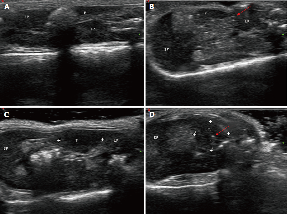
Figure 5 Evaluation of therapeutic effects of irreversible electroporation on tumors in vivo by ultrasound.
A: A pre-irreversible electroporation ultrasound image showing the normal pancreatic parenchyma; B: The ablation zone showed hyperechoic signals with a comet tail sign in the normal pancreatic tissue (arrow); C: White dots indicate the region of the tumor; D: Ultrasound image showing that the irreversible electroporation ablation zone in the tumor tissue became hyperechoic (arrow). SP: Spleen; LK: Left kidney; P: Pancreas; T: Tumor.
- Citation: Su JJ, Xu K, Wang PF, Zhang HY, Chen YL. Histological analysis of human pancreatic carcinoma following irreversible electroporation in a nude mouse model. World J Gastrointest Oncol 2018; 10(12): 476-486
- URL: https://www.wjgnet.com/1948-5204/full/v10/i12/476.htm
- DOI: https://dx.doi.org/10.4251/wjgo.v10.i12.476


