Copyright
©The Author(s) 2018.
World J Gastrointest Oncol. Dec 15, 2018; 10(12): 476-486
Published online Dec 15, 2018. doi: 10.4251/wjgo.v10.i12.476
Published online Dec 15, 2018. doi: 10.4251/wjgo.v10.i12.476
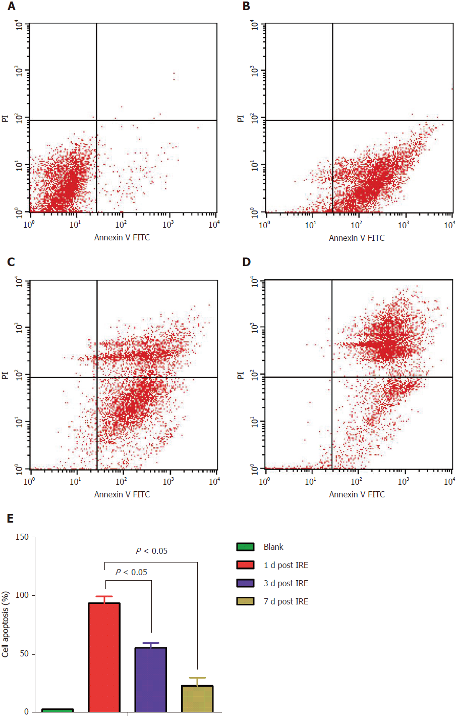
Figure 4 Apoptosis assay of mouse spleen cells before and after irreversible electroporation treatment using double-staining with annexin V-fluorescein isothiocyanate/propidium iodide.
Apoptosis was quantified by flow cytometry. A: Control group; B: 1 d post-IRE; C: 3 d post-IRE; D: 7 d post-IRE; E: Percentages of apoptotic cells before and after the IRE intervention. Data are mean ± SD (n = 8). IRE: irreversible electroporation.
- Citation: Su JJ, Xu K, Wang PF, Zhang HY, Chen YL. Histological analysis of human pancreatic carcinoma following irreversible electroporation in a nude mouse model. World J Gastrointest Oncol 2018; 10(12): 476-486
- URL: https://www.wjgnet.com/1948-5204/full/v10/i12/476.htm
- DOI: https://dx.doi.org/10.4251/wjgo.v10.i12.476


