Copyright
©The Author(s) 2018.
World J Gastrointest Oncol. Dec 15, 2018; 10(12): 476-486
Published online Dec 15, 2018. doi: 10.4251/wjgo.v10.i12.476
Published online Dec 15, 2018. doi: 10.4251/wjgo.v10.i12.476
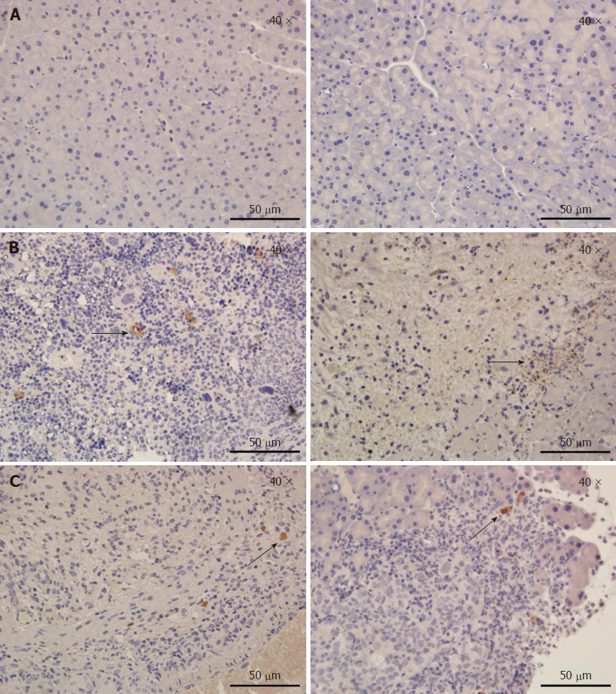
Figure 3 Tumor tissue sections at 1 d and 7 d after the irreversible electroporation treatment were stained for Ki67 and cleaved caspase-3.
A: A representative immunohistochemistry image in an untreated pancreatic parenchyma; B: In tumor tissues, extensive caspase-3 activation was observed on the first postoperative day. Irreversible electroporation significantly increased cell proliferation (Ki67 staining) at 1 d post-treatment, but cell proliferation was decreased at 7 d post-treatment (arrows); C: Limited caspase-3 staining at 7 d post-irreversible electroporation treatment was found in treated tumors, while most of the viable tumor tissues showed no caspase-3 activation. The slides were imaged at 400 × by light microscopy.
- Citation: Su JJ, Xu K, Wang PF, Zhang HY, Chen YL. Histological analysis of human pancreatic carcinoma following irreversible electroporation in a nude mouse model. World J Gastrointest Oncol 2018; 10(12): 476-486
- URL: https://www.wjgnet.com/1948-5204/full/v10/i12/476.htm
- DOI: https://dx.doi.org/10.4251/wjgo.v10.i12.476


