Copyright
©The Author(s) 2018.
World J Gastrointest Oncol. Nov 15, 2018; 10(11): 439-448
Published online Nov 15, 2018. doi: 10.4251/wjgo.v10.i11.439
Published online Nov 15, 2018. doi: 10.4251/wjgo.v10.i11.439
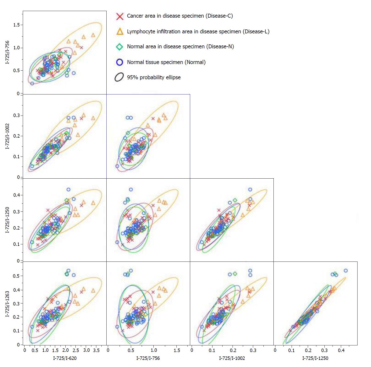
Figure 4 Biaxial distribution of the Raman scattering intensity ratio with the intensity of wavenumber 725 cm-1 as the denominator.
- Citation: Ikeda H, Ito H, Hikita M, Yamaguchi N, Uragami N, Yokoyama N, Hirota Y, Kushima M, Ajioka Y, Inoue H. Raman spectroscopy for the diagnosis of unlabeled and unstained histopathological tissue specimens. World J Gastrointest Oncol 2018; 10(11): 439-448
- URL: https://www.wjgnet.com/1948-5204/full/v10/i11/439.htm
- DOI: https://dx.doi.org/10.4251/wjgo.v10.i11.439


