Copyright
©The Author(s) 2018.
World J Gastrointest Oncol. Nov 15, 2018; 10(11): 439-448
Published online Nov 15, 2018. doi: 10.4251/wjgo.v10.i11.439
Published online Nov 15, 2018. doi: 10.4251/wjgo.v10.i11.439
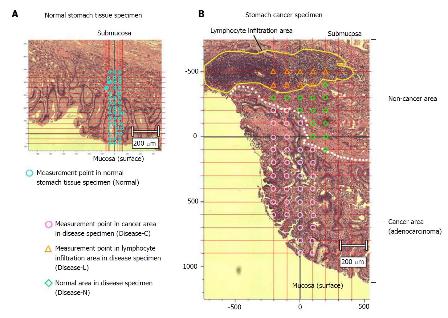
Figure 2 Measured points in the stomach cancer and normal tissue specimens.
A: Normal stomach tissue specimen; B: Stomach cancer specimen. We established the conditions for laser output and laser irradiation time on a marginal part of an unstained tissue specimen that included both gastric cancer lesion and non-lesion areas. To prevent tissue degeneration, we reduced the laser power as much as possible, while maintaining detection of the Raman spectrum. Optimal measurement conditions were established as a laser output of 1.7 mW and an irradiation time of 10 s. We measured the tissue specimens at regular intervals from the mucous membrane to the submucosal layer.
- Citation: Ikeda H, Ito H, Hikita M, Yamaguchi N, Uragami N, Yokoyama N, Hirota Y, Kushima M, Ajioka Y, Inoue H. Raman spectroscopy for the diagnosis of unlabeled and unstained histopathological tissue specimens. World J Gastrointest Oncol 2018; 10(11): 439-448
- URL: https://www.wjgnet.com/1948-5204/full/v10/i11/439.htm
- DOI: https://dx.doi.org/10.4251/wjgo.v10.i11.439


