Copyright
©The Author(s) 2018.
World J Gastrointest Oncol. Nov 15, 2018; 10(11): 439-448
Published online Nov 15, 2018. doi: 10.4251/wjgo.v10.i11.439
Published online Nov 15, 2018. doi: 10.4251/wjgo.v10.i11.439
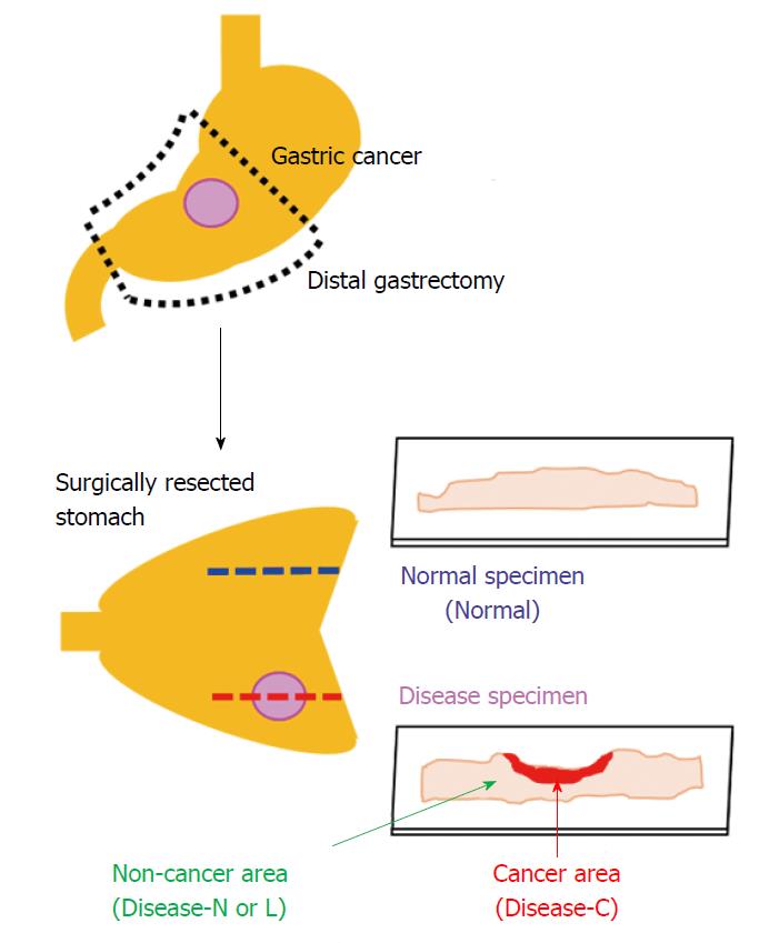
Figure 1 Two consecutive tissue specimens from areas with and without stomach cancer lesions.
Each tissue specimen was sliced to a 3-μm thickness with a microtome and attached to a 1-mm-thick low-autofluorescence slide (SUPER FROST, Matsunami Glass Ind., Ltd, Tokyo, Japan). A thin cover glass (NEO microscope cover glass, Matsunami Glass Ind., Ltd., Tokyo, Japan) was placed on the tissue. Sections were deparaffinized by sequential immersion in xylene, ethanol, and water. One of the two tissue specimens was stained with hematoxylin and eosin and used as a reference for laser irradiation positioning by the spectroscopic method. Another tissue specimen was left unstained and used for analysis by Raman spectroscopy. We acquired the Raman spectrum of the cancer area (Disease-C), non-cancerous lymphocytes infiltration area (Disease-L), non-cancerous normal area (Disease-N) in the stomach cancer specimen, and normal stomach tissue specimen (Normal).
- Citation: Ikeda H, Ito H, Hikita M, Yamaguchi N, Uragami N, Yokoyama N, Hirota Y, Kushima M, Ajioka Y, Inoue H. Raman spectroscopy for the diagnosis of unlabeled and unstained histopathological tissue specimens. World J Gastrointest Oncol 2018; 10(11): 439-448
- URL: https://www.wjgnet.com/1948-5204/full/v10/i11/439.htm
- DOI: https://dx.doi.org/10.4251/wjgo.v10.i11.439


