Copyright
©The Author(s) 2018.
World J Gastrointest Oncol. Nov 15, 2018; 10(11): 381-397
Published online Nov 15, 2018. doi: 10.4251/wjgo.v10.i11.381
Published online Nov 15, 2018. doi: 10.4251/wjgo.v10.i11.381
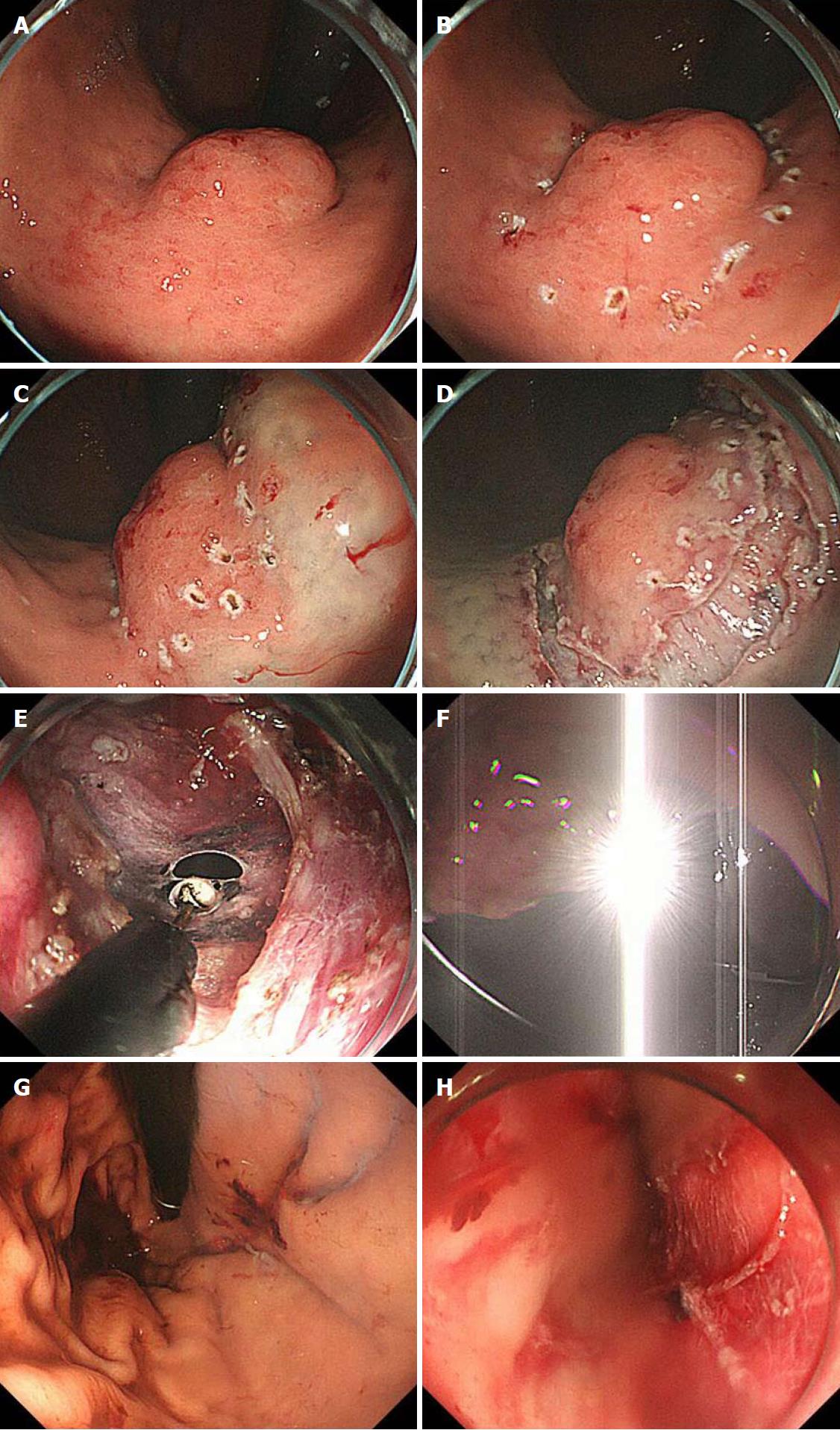
Figure 4 Intraoperative endoscopic view of laparoscopic and endoscopic cooperative surgery.
A: First, the location of the tumor is confirmed; B: The periphery of the tumor is marked using argon plasma coagulation as close as possible to the tumor edge; C: Glycerin mixed with indigo blue is injected into the submucosal layer; D: The whole circumference of the marked area is cut using an insulation-tipped diathermic knife; E: An intentional perforation is made; F: The laparoscopic light is too dazzling for the endoscopic side; G: Intraluminal view after suturing. The absence of stenosis and malformation is confirmed; H: Esophageal mucosa injury by the plastic bag during specimen removal.
- Citation: Aisu Y, Yasukawa D, Kimura Y, Hori T. Laparoscopic and endoscopic cooperative surgery for gastric tumors: Perspective for actual practice and oncological benefits. World J Gastrointest Oncol 2018; 10(11): 381-397
- URL: https://www.wjgnet.com/1948-5204/full/v10/i11/381.htm
- DOI: https://dx.doi.org/10.4251/wjgo.v10.i11.381


