Copyright
©The Author(s) 2018.
World J Gastrointest Oncol. Nov 15, 2018; 10(11): 370-380
Published online Nov 15, 2018. doi: 10.4251/wjgo.v10.i11.370
Published online Nov 15, 2018. doi: 10.4251/wjgo.v10.i11.370
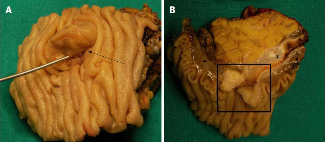
Figure 1 A classic example of the macroscopic appearance of a case of ampulla of Vater carcinoma.
A: The ampullary area is markedly enlarged (black arrow); B: On the section surface, the ampulla of Vater carcinoma (black box), the adjacent duodenal wall (black arrow) and bile duct (asterisk) are clearly visible.
- Citation: Pea A, Riva G, Bernasconi R, Sereni E, Lawlor RT, Scarpa A, Luchini C. Ampulla of Vater carcinoma: Molecular landscape and clinical implications. World J Gastrointest Oncol 2018; 10(11): 370-380
- URL: https://www.wjgnet.com/1948-5204/full/v10/i11/370.htm
- DOI: https://dx.doi.org/10.4251/wjgo.v10.i11.370


