Copyright
©The Author(s) 2017.
World J Gastrointest Endosc. Sep 16, 2017; 9(9): 494-498
Published online Sep 16, 2017. doi: 10.4253/wjge.v9.i9.494
Published online Sep 16, 2017. doi: 10.4253/wjge.v9.i9.494
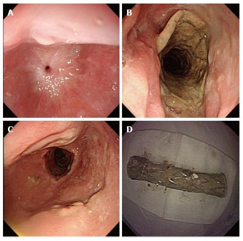
Figure 3 Endoscopic appearances of the esophageal stricture after all the stents were removed.
A: The esophageal stricture located 25 cm from the incisor teeth was worsened 5 wk after the stents were removed, making it difficult to pass the gastroscope; B: A fully covered SEMS was placed and left in place for 4 wk; C: The stricture was significantly wider after the removal of (D) the fully covered SEMS. SEMS: Self-expanding metal stent.
- Citation: Liu XQ, Zhou M, Shi WX, Qi YY, Liu H, Li B, Xu HW. Successful endoscopic removal of three embedded esophageal self-expanding metal stents. World J Gastrointest Endosc 2017; 9(9): 494-498
- URL: https://www.wjgnet.com/1948-5190/full/v9/i9/494.htm
- DOI: https://dx.doi.org/10.4253/wjge.v9.i9.494


