Copyright
©The Author(s) 2017.
World J Gastrointest Endosc. Sep 16, 2017; 9(9): 494-498
Published online Sep 16, 2017. doi: 10.4253/wjge.v9.i9.494
Published online Sep 16, 2017. doi: 10.4253/wjge.v9.i9.494
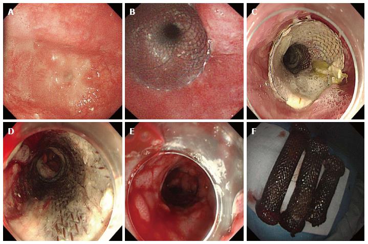
Figure 2 Gastroscope images of esophagus and stents.
Gastroscopy showed (A) an esophageal stricture beginning 25 cm from the incisor teeth; B: The fourth stent was placed and (C) remained in place for 4 wk; D: The previous stents became visible after the removal of the fourth stent; E: Outcomes after the removal of all stents were diffuse, but minor bleeding and mucosal tears, and the imprint of the stent mesh was visible in the esophageal mucosa; F: The three original stents were completely retrieved.
- Citation: Liu XQ, Zhou M, Shi WX, Qi YY, Liu H, Li B, Xu HW. Successful endoscopic removal of three embedded esophageal self-expanding metal stents. World J Gastrointest Endosc 2017; 9(9): 494-498
- URL: https://www.wjgnet.com/1948-5190/full/v9/i9/494.htm
- DOI: https://dx.doi.org/10.4253/wjge.v9.i9.494


