Copyright
©The Author(s) 2017.
World J Gastrointest Endosc. Sep 16, 2017; 9(9): 438-447
Published online Sep 16, 2017. doi: 10.4253/wjge.v9.i9.438
Published online Sep 16, 2017. doi: 10.4253/wjge.v9.i9.438
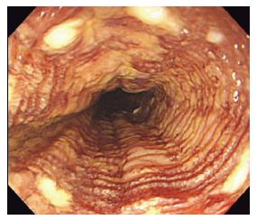
Figure 2 “Tatami sign” is commonly seen after iodine staining.
It is characterized by regular, fine circular folds of the Lugol’s unstained area. This is typically seen when lesions are confined to the muscularis mucosal.
- Citation: Shimamura Y, Ikeya T, Marcon N, Mosko JD. Endoscopic diagnosis and treatment of early esophageal squamous neoplasia. World J Gastrointest Endosc 2017; 9(9): 438-447
- URL: https://www.wjgnet.com/1948-5190/full/v9/i9/438.htm
- DOI: https://dx.doi.org/10.4253/wjge.v9.i9.438


