Copyright
©The Author(s) 2017.
World J Gastrointest Endosc. Aug 16, 2017; 9(8): 417-424
Published online Aug 16, 2017. doi: 10.4253/wjge.v9.i8.417
Published online Aug 16, 2017. doi: 10.4253/wjge.v9.i8.417
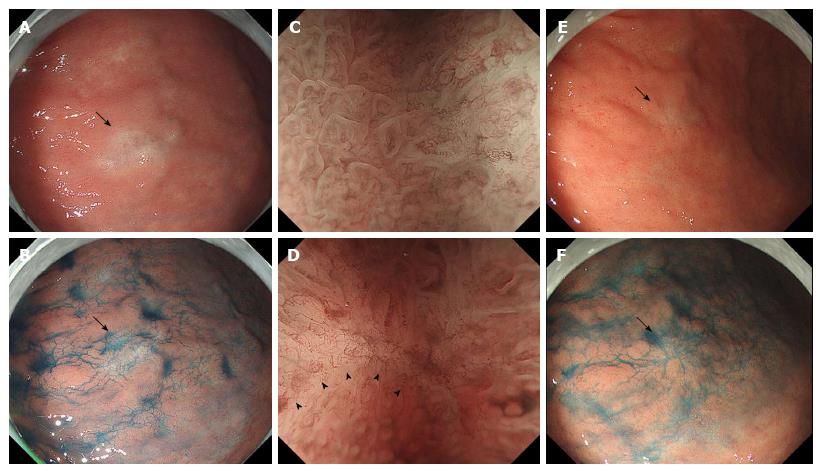
Figure 1 Endoscopic imaging of the antral lesion before and after the eradication.
A: An endoscopic image before the eradication shows a 13-mm whitish/slightly brownish lesion (arrow) on the lesser curvature of the antrum; B: Indigo carmine dye spray reveals that the antral lesion (arrow) is almost flat; C: Magnification endoscopy with narrow-band imaging shows the lesion (C, right side) exhibits loss or irregularity of the microsurface pattern and irregular microvascular proliferation compared to the background mucosa (C, left side). A demarcation line (D, arrowheads) is seen at the periphery of the lesion. E-F: Endoscopic images after the eradication therapy exhibit that the mucosal lesion (arrow) decreases in size to 7 mm in diameter (E) and preserves the gross appearance with indigo carmine dye spray (F).
- Citation: Yorita K, Iwasaki T, Uchita K, Kuroda N, Kojima K, Iwamura S, Tsutsumi Y, Ohno A, Kataoka H. Russell body gastritis with Dutcher bodies evaluated using magnification endoscopy. World J Gastrointest Endosc 2017; 9(8): 417-424
- URL: https://www.wjgnet.com/1948-5190/full/v9/i8/417.htm
- DOI: https://dx.doi.org/10.4253/wjge.v9.i8.417


