Copyright
©The Author(s) 2017.
World J Gastrointest Endosc. Jul 16, 2017; 9(7): 319-326
Published online Jul 16, 2017. doi: 10.4253/wjge.v9.i7.319
Published online Jul 16, 2017. doi: 10.4253/wjge.v9.i7.319
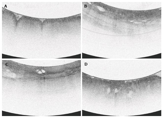
Figure 3 Volumetric laser endomicroscopy imaging snapshots.
A: Normal gastric cardia with gastric rugae and gastric pit architecture; B: Inflamed gastric cardia with loss of gastric pit architecture and anomalous glands; C: Low grade dysplasia with loss of gastric pit architecture, heterogeneous scattering, anomalous septated gland; D: High grade dysplasia with irregular surface and anomalous glands.
- Citation: Gupta N, Siddiqui U, Waxman I, Chapman C, Koons A, Valuckaite V, Xiao SY, Setia N, Hart J, Konda V. Use of volumetric laser endomicroscopy for dysplasia detection at the gastroesophageal junction and gastric cardia. World J Gastrointest Endosc 2017; 9(7): 319-326
- URL: https://www.wjgnet.com/1948-5190/full/v9/i7/319.htm
- DOI: https://dx.doi.org/10.4253/wjge.v9.i7.319


