Copyright
©The Author(s) 2017.
World J Gastrointest Endosc. Jun 16, 2017; 9(6): 243-254
Published online Jun 16, 2017. doi: 10.4253/wjge.v9.i6.243
Published online Jun 16, 2017. doi: 10.4253/wjge.v9.i6.243
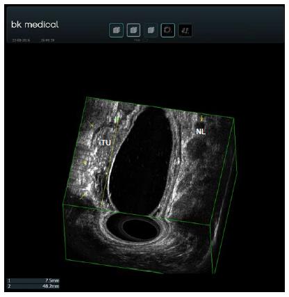
Figure 6 Rectal adenocarcinoma staging by 3D endoscopic ultrasound T1 N1.
The yellow arrows on the left show the muscularis propria. The tumor invades up to the submucosa. A white submucosa plane can be seen between the tumor (TU) and the muscularis propria. The yellow arrow on the right shows a round lymph node. The 3D image was obtained using a transanal rigid probe with an ultrasound from bk medical.
- Citation: Valero M, Robles-Medranda C. Endoscopic ultrasound in oncology: An update of clinical applications in the gastrointestinal tract. World J Gastrointest Endosc 2017; 9(6): 243-254
- URL: https://www.wjgnet.com/1948-5190/full/v9/i6/243.htm
- DOI: https://dx.doi.org/10.4253/wjge.v9.i6.243


