Copyright
©The Author(s) 2017.
World J Gastrointest Endosc. Jun 16, 2017; 9(6): 243-254
Published online Jun 16, 2017. doi: 10.4253/wjge.v9.i6.243
Published online Jun 16, 2017. doi: 10.4253/wjge.v9.i6.243
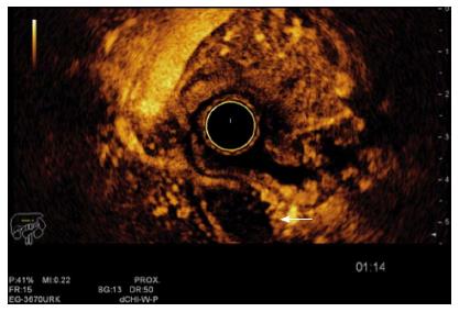
Figure 5 The same lesion presented in Figure 3 being evaluated by contrast enhanced ultrasonography.
The white arrow shows the lymph node with no enhancement after the contrast application, which suggests malignancy. The endoscopic ultrasound-contrast enhancement was done using a Hitachi-Avius console with a radial scope EG-3630URK (from Pentax Medical) and a Sonovue contrast agent (from Bracco).
- Citation: Valero M, Robles-Medranda C. Endoscopic ultrasound in oncology: An update of clinical applications in the gastrointestinal tract. World J Gastrointest Endosc 2017; 9(6): 243-254
- URL: https://www.wjgnet.com/1948-5190/full/v9/i6/243.htm
- DOI: https://dx.doi.org/10.4253/wjge.v9.i6.243


