Copyright
©The Author(s) 2017.
World J Gastrointest Endosc. May 16, 2017; 9(5): 211-219
Published online May 16, 2017. doi: 10.4253/wjge.v9.i5.211
Published online May 16, 2017. doi: 10.4253/wjge.v9.i5.211
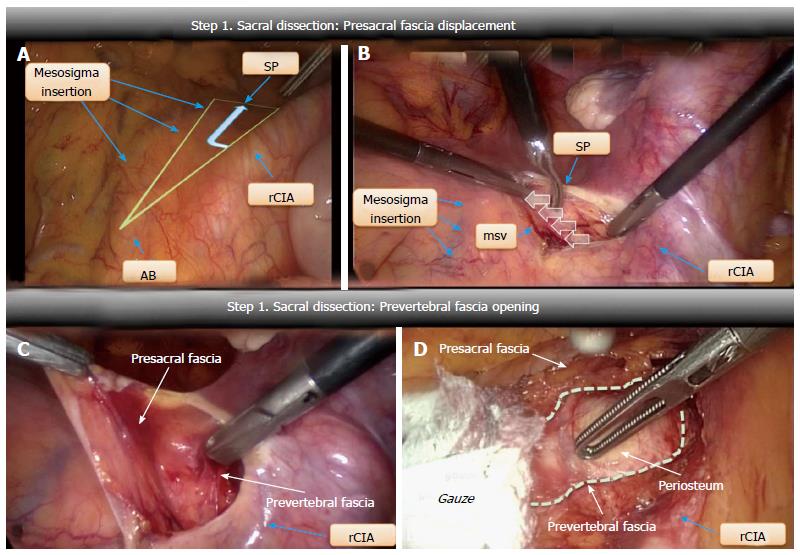
Figure 2 Step 1: Presacral fascia displacement, prevertebral fascia opening.
A: The imaginary outline of the “dissection triangle” on the right lumbosacral spine between the four anatomical landmarks; B: Presacral fascia displacement not beyond the middle sacral vein (msv); C: Presacral fascia and prevertebral fascia; D: Denuded periosteum after prevertebral fascia opening. rCIA: Right common iliac artery; SP: Sacral promontory; AB: Aortic bifurcation.
- Citation: Cosma S, Petruzzelli P, Danese S, Benedetto C. Nerve preserving vs standard laparoscopic sacropexy: Postoperative bowel function. World J Gastrointest Endosc 2017; 9(5): 211-219
- URL: https://www.wjgnet.com/1948-5190/full/v9/i5/211.htm
- DOI: https://dx.doi.org/10.4253/wjge.v9.i5.211


