Copyright
©The Author(s) 2017.
World J Gastrointest Endosc. Mar 16, 2017; 9(3): 139-144
Published online Mar 16, 2017. doi: 10.4253/wjge.v9.i3.139
Published online Mar 16, 2017. doi: 10.4253/wjge.v9.i3.139
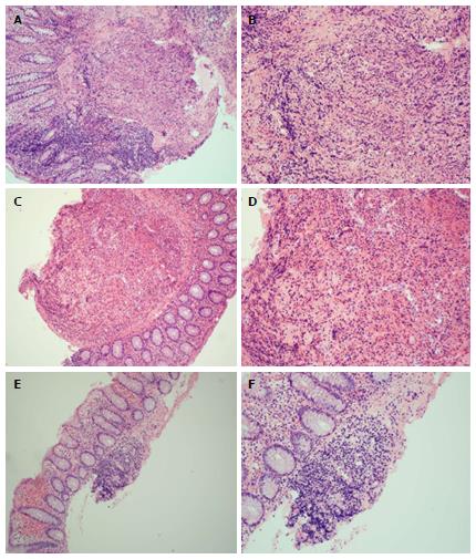
Figure 3 Histology of colonic samples consistent with Langerhans cell histiocytosis.
Grossly, the colon biopsies were each received as small fragments of tissue, where no tissue defects could be determined due to the size of the specimens received. Microscopically, the cecum (A and B, 10 × and 20 ×, respectively), descending colon (C and D, 10 × and 40 ×, respectively), and transverse colon (E and F, 10 × and 20 ×, respectively) each show nodules that are similar in morphology to the appendix and (although not shown here) share the same staining patterns (CD1a, S-100, and langerin reactivity). Again, electron microscopy was unsuccessful in highlighting the Birbeck bodies due to formalin fixation.
- Citation: Karimzada MM, Matthews MN, French SW, DeUgarte D, Kim DY. Langerhans cell histiocytosis masquerading as acute appendicitis: Case report and review. World J Gastrointest Endosc 2017; 9(3): 139-144
- URL: https://www.wjgnet.com/1948-5190/full/v9/i3/139.htm
- DOI: https://dx.doi.org/10.4253/wjge.v9.i3.139


