Copyright
©The Author(s) 2017.
World J Gastrointest Endosc. Mar 16, 2017; 9(3): 139-144
Published online Mar 16, 2017. doi: 10.4253/wjge.v9.i3.139
Published online Mar 16, 2017. doi: 10.4253/wjge.v9.i3.139
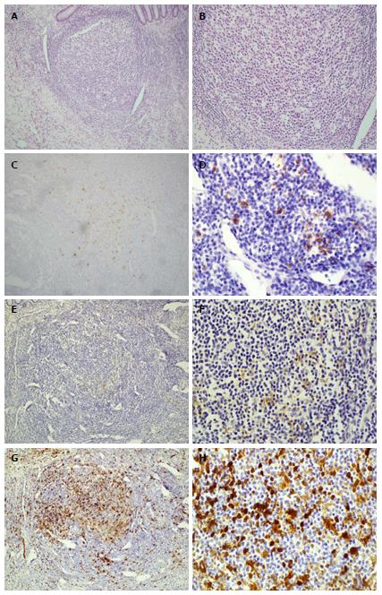
Figure 1 Histology and immunohistochemical stains of appendix confirming diagnosis of Langerhans cell histiocytosis.
The appendix shows an overgrowth of histiocytes in the lymphoid aggregates at 10 × (A) and 40 × (B) magnifications of H and E stains; while the electron microscopy images did not show Birbeck bodies due to previous fixation, the Langerin stain (CD207) demonstrates their presence in the areas of concern at 10 × (C) and 40 × (D) magnification. CD1a stains at 10 × (E) and 40 × (F); S-100 stains at 10 × (G) and 40 × (H).
- Citation: Karimzada MM, Matthews MN, French SW, DeUgarte D, Kim DY. Langerhans cell histiocytosis masquerading as acute appendicitis: Case report and review. World J Gastrointest Endosc 2017; 9(3): 139-144
- URL: https://www.wjgnet.com/1948-5190/full/v9/i3/139.htm
- DOI: https://dx.doi.org/10.4253/wjge.v9.i3.139


