Copyright
©The Author(s) 2017.
World J Gastrointest Endosc. Feb 16, 2017; 9(2): 99-104
Published online Feb 16, 2017. doi: 10.4253/wjge.v9.i2.99
Published online Feb 16, 2017. doi: 10.4253/wjge.v9.i2.99
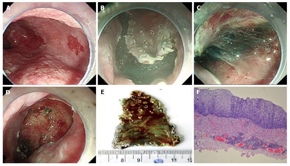
Figure 4 Endoscopic submucosal dissection procedure and pathological examination.
A-D: Marking around the lesion using Dual knife; submucosal injection of 10 mL saline with 0.3% indigo carmine and 1:100000 epinephrine; cutting open the mucosa; the submucosa was stripped and the lesion was completely resected; E, F: Pathological examination of the resected specimen revealed high-grade intraepithelial neoplasia with a component of scattered low-grade intraepithelial neoplasia. Both of the lateral and vertical margins were negative of tumor.
- Citation: Shi S, Fu K, Dong XQ, Hao YJ, Li SL. Combination of concurrent endoscopic submucosal dissection and modified peroral endoscopic myotomy for an achalasia patient with synchronous early esophageal neoplasms. World J Gastrointest Endosc 2017; 9(2): 99-104
- URL: https://www.wjgnet.com/1948-5190/full/v9/i2/99.htm
- DOI: https://dx.doi.org/10.4253/wjge.v9.i2.99


