Copyright
©The Author(s) 2017.
World J Gastrointest Endosc. Feb 16, 2017; 9(2): 99-104
Published online Feb 16, 2017. doi: 10.4253/wjge.v9.i2.99
Published online Feb 16, 2017. doi: 10.4253/wjge.v9.i2.99
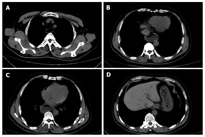
Figure 1 Chest computed tomography examination showed that the esophageal cavity was obviously expanded (A-C); large amount of fluid retention was seen in the lumen (D).
The cardiac muscle layer was significantly thickened.
- Citation: Shi S, Fu K, Dong XQ, Hao YJ, Li SL. Combination of concurrent endoscopic submucosal dissection and modified peroral endoscopic myotomy for an achalasia patient with synchronous early esophageal neoplasms. World J Gastrointest Endosc 2017; 9(2): 99-104
- URL: https://www.wjgnet.com/1948-5190/full/v9/i2/99.htm
- DOI: https://dx.doi.org/10.4253/wjge.v9.i2.99


