Copyright
©The Author(s) 2016.
World J Gastrointest Endosc. Apr 25, 2016; 8(8): 374-377
Published online Apr 25, 2016. doi: 10.4253/wjge.v8.i8.374
Published online Apr 25, 2016. doi: 10.4253/wjge.v8.i8.374
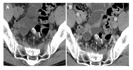
Figure 3 Un-enhanced and intravenous contrast-enhanced computed tomography scans of the abdomen show a slightly hyperdense mass in the rectal wall without contract enhancement (white arrow).
A: Unenhanced CT; B: Contrast enhanced CT. CT: Computed tomography.
- Citation: Morimoto M, Koinuma K, Lefor AK, Horie H, Ito H, Sata N, Hayashi Y, Sunada K, Yamamoto H. Diagnosis of a submucosal mass at the staple line after sigmoid colon cancer resection by endoscopic cutting-mucosa biopsy. World J Gastrointest Endosc 2016; 8(8): 374-377
- URL: https://www.wjgnet.com/1948-5190/full/v8/i8/374.htm
- DOI: https://dx.doi.org/10.4253/wjge.v8.i8.374


