Copyright
©The Author(s) 2016.
World J Gastrointest Endosc. Feb 25, 2016; 8(4): 244-251
Published online Feb 25, 2016. doi: 10.4253/wjge.v8.i4.244
Published online Feb 25, 2016. doi: 10.4253/wjge.v8.i4.244
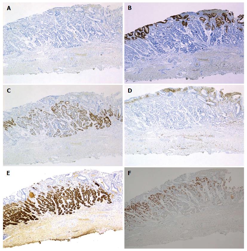
Figure 3 Immunohistochemical staining: Case 1.
Tumor cells were diffusely positive for MUC6 (C) and pepsinogen-I (E), partially positive for H+/K+-ATPase in scattered locations around the tumor margin (F), negative staining for MUC2 (A), MUC5AC (B) and CD10 (D). Magnification: A-F (low-power view × 40). MUC: Mucin.
- Citation: Tohda G, Osawa T, Asada Y, Dochin M, Terahata S. Gastric adenocarcinoma of fundic gland type: Endoscopic and clinicopathological features. World J Gastrointest Endosc 2016; 8(4): 244-251
- URL: https://www.wjgnet.com/1948-5190/full/v8/i4/244.htm
- DOI: https://dx.doi.org/10.4253/wjge.v8.i4.244


