Copyright
©The Author(s) 2016.
World J Gastrointest Endosc. Feb 10, 2016; 8(3): 192-197
Published online Feb 10, 2016. doi: 10.4253/wjge.v8.i3.192
Published online Feb 10, 2016. doi: 10.4253/wjge.v8.i3.192
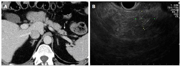
Figure 4 Twenty-four months follow-up.
A: Computed tomography scan showing absence of hypervascular tissue around a small hypodense area (white arrow); B: Endoscopic ultrasound scan of the pancreatic tail demonstrating poorly defined hyperechoic tissue (fibrosis) with posterior shadow (caliper).
- Citation: Armellini E, Crinò SF, Ballarè M, Pallio S, Occhipinti P. Endoscopic ultrasound-guided ethanol ablation of pancreatic neuroendocrine tumours: A case study and literature review. World J Gastrointest Endosc 2016; 8(3): 192-197
- URL: https://www.wjgnet.com/1948-5190/full/v8/i3/192.htm
- DOI: https://dx.doi.org/10.4253/wjge.v8.i3.192


