Copyright
©The Author(s) 2016.
World J Gastrointest Endosc. Jan 25, 2016; 8(2): 67-76
Published online Jan 25, 2016. doi: 10.4253/wjge.v8.i2.67
Published online Jan 25, 2016. doi: 10.4253/wjge.v8.i2.67
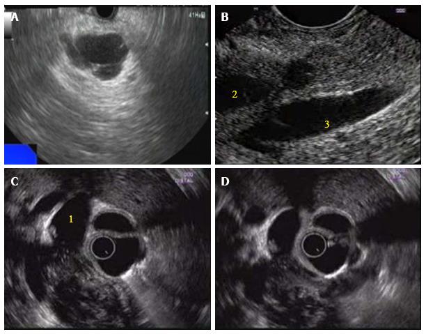
Figure 1 Endoscopic ultrasound appearance of mass lesions in pancreas.
A: Serous cystic neoplasm of head of pancreas (HOP); B: Neuroendocrine tumor of head of pancreas with dilated pancreatic duct (2) and adjacent portal vein (3); C: Carcinoma HOP with loss of fat planes with confluence of superior mesenteric vein (SMV) and portal vein and dilated common bile duct (1); D: Carcinoma HOP with common bile duct and SMV infiltration.
- Citation: Puri R, Manrai M, Thandassery RB, Alfadda AA. Endoscopic ultrasound in the diagnosis and management of carcinoma pancreas. World J Gastrointest Endosc 2016; 8(2): 67-76
- URL: https://www.wjgnet.com/1948-5190/full/v8/i2/67.htm
- DOI: https://dx.doi.org/10.4253/wjge.v8.i2.67


