Copyright
©The Author(s) 2016.
World J Gastrointest Endosc. Jan 25, 2016; 8(2): 56-66
Published online Jan 25, 2016. doi: 10.4253/wjge.v8.i2.56
Published online Jan 25, 2016. doi: 10.4253/wjge.v8.i2.56
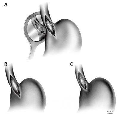
Figure 2 Esophageal myotomies.
A: The original Heller myotomy, consisting of both anterior and posterior disruption of esophageal fibers; B: The most commonly performed Heller myotomy, with extension onto the stomach for 2-3 cm; C: Heller myotomy with minimal extension onto the stomach.
- Citation: Pandian TK, Naik ND, Fahy AS, Arghami A, Farley DR, Ishitani MB, Moir CR. Laparoscopic esophagomyotomy for achalasia in children: A review. World J Gastrointest Endosc 2016; 8(2): 56-66
- URL: https://www.wjgnet.com/1948-5190/full/v8/i2/56.htm
- DOI: https://dx.doi.org/10.4253/wjge.v8.i2.56


