Copyright
©The Author(s) 2016.
World J Gastrointest Endosc. Nov 16, 2016; 8(19): 690-696
Published online Nov 16, 2016. doi: 10.4253/wjge.v8.i19.690
Published online Nov 16, 2016. doi: 10.4253/wjge.v8.i19.690
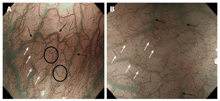
Figure 3 High magnifying narrow band imaging image of normal esophageal mucosa (luminal side).
A: Soft pressure of the endoscope distal attachment (“hood”) onto the mucosal surface demonstrates SECN, hard pressure onto the mucosa compresses horizontal vessels, allowing clear observation of IPCLs; B: In the circle the SECN located at the top layer of lamina propria mucosae, just beneath the epithelium. The black arrows indicate the branching vessels into the lower lamina propria; white arrows indicate the IPCL located in the epithelial papilla, which is a projection of lamina propria mucosae into the epithelium. SECN: Sub-epithelial capillary network; IPCL: Intrapapillary capillary loop.
- Citation: Maselli R, Inoue H, Ikeda H, Onimaru M, Yoshida A, Santi EG, Sato H, Hayee B, Kudo SE. Microvasculature of the esophagus and gastroesophageal junction: Lesson learned from submucosal endoscopy. World J Gastrointest Endosc 2016; 8(19): 690-696
- URL: https://www.wjgnet.com/1948-5190/full/v8/i19/690.htm
- DOI: https://dx.doi.org/10.4253/wjge.v8.i19.690


