Copyright
©The Author(s) 2016.
World J Gastrointest Endosc. Oct 16, 2016; 8(18): 669-673
Published online Oct 16, 2016. doi: 10.4253/wjge.v8.i18.669
Published online Oct 16, 2016. doi: 10.4253/wjge.v8.i18.669
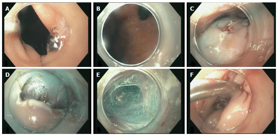
Figure 1 Endoscopic pictures from the first per oral endoscopic myotomy attempt showing: Gastroesophageal junction (A), cardia before myotomy (B), mucosotomy site (C), initial dissection site (D), creating the submucosal tunnel (E), and closure of mucosotomy (F).
- Citation: Wehbeh AN, Mekaroonkamol P, Cai Q. Same site submucosal tunneling for a repeat per oral endoscopic myotomy: A safe and feasible option. World J Gastrointest Endosc 2016; 8(18): 669-673
- URL: https://www.wjgnet.com/1948-5190/full/v8/i18/669.htm
- DOI: https://dx.doi.org/10.4253/wjge.v8.i18.669


