Copyright
©The Author(s) 2016.
World J Gastrointest Endosc. Oct 16, 2016; 8(18): 653-662
Published online Oct 16, 2016. doi: 10.4253/wjge.v8.i18.653
Published online Oct 16, 2016. doi: 10.4253/wjge.v8.i18.653
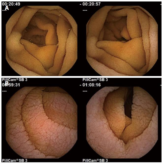
Figure 1 Normal (A) vs untreated celiac patient images (B).
Note the presence of mucosal folds, a mottled appearance, and fissuring, in the images from untreated celiac patients (lower).
- Citation: Ciaccio EJ, Bhagat G, Lewis SK, Green PH. Recommendations to quantify villous atrophy in video capsule endoscopy images of celiac disease patients. World J Gastrointest Endosc 2016; 8(18): 653-662
- URL: https://www.wjgnet.com/1948-5190/full/v8/i18/653.htm
- DOI: https://dx.doi.org/10.4253/wjge.v8.i18.653


