Copyright
©The Author(s) 2016.
World J Gastrointest Endosc. Aug 25, 2016; 8(16): 558-567
Published online Aug 25, 2016. doi: 10.4253/wjge.v8.i16.558
Published online Aug 25, 2016. doi: 10.4253/wjge.v8.i16.558
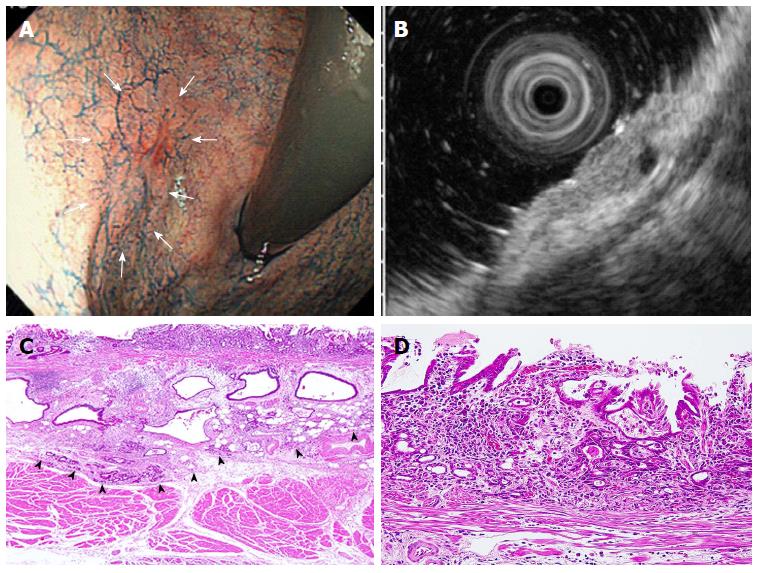
Figure 2 Case diagnosed correctly by endoscopic ultrasonography but misdiagnosed by endoscopy.
A: Chromoendoscopy shows a reddish and smooth surface in a shallow depressed lesion diagnosed as M/SM1 (arrows). Histologically, the biopsy sample indicated a moderately to poorly differentiated adenocarcinoma; B: EUS image showing that a hypoechoic mass invaded the submucosal layer (sonographic layer 3). This lesion was diagnosed as SM2; C: Histology revealed that undifferentiated type adenocarcinoma massively invaded the submucosal layer (arrowheads); D: Moderately to poorly differentiated adenocarcinoma cells were observed in the gastric mucosae (× 200). EUS: Endoscopic ultrasonography.
- Citation: Watari J, Ueyama S, Tomita T, Ikehara H, Hori K, Hara K, Yamasaki T, Okugawa T, Kondo T, Kono T, Tozawa K, Oshima T, Fukui H, Miwa H. What types of early gastric cancer are indicated for endoscopic ultrasonography staging of invasion depth? World J Gastrointest Endosc 2016; 8(16): 558-567
- URL: https://www.wjgnet.com/1948-5190/full/v8/i16/558.htm
- DOI: https://dx.doi.org/10.4253/wjge.v8.i16.558


