Copyright
©The Author(s) 2015.
World J Gastrointest Endosc. Jul 25, 2015; 7(9): 872-880
Published online Jul 25, 2015. doi: 10.4253/wjge.v7.i9.872
Published online Jul 25, 2015. doi: 10.4253/wjge.v7.i9.872
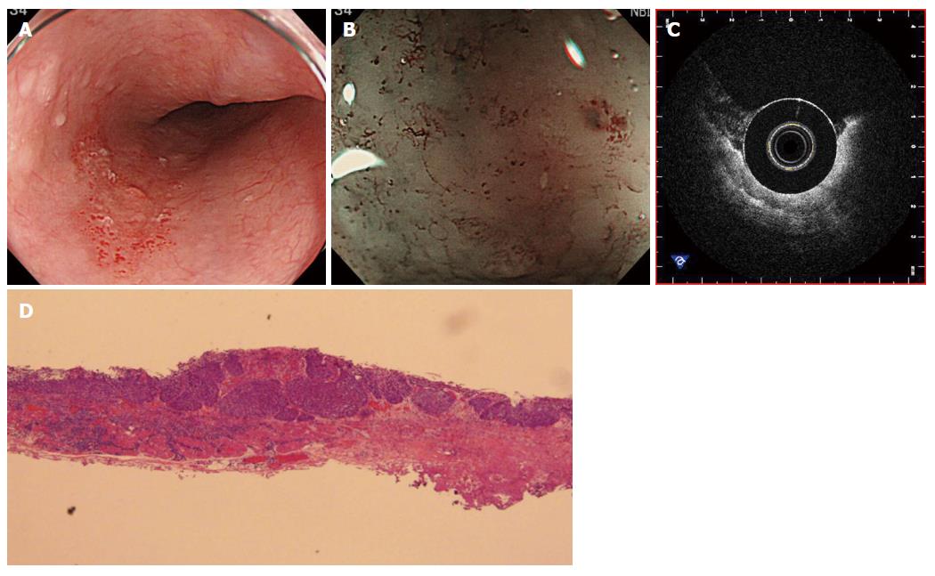
Figure 1 Representative images of superficial esophageal squamous cell carcinoma (0-IIc) with the recurrence after chemo-radiation therapy.
A 71-year-old patient suffered from the recurrence of esophageal squamous cell carcinoma after chemo-radiation therapy. A: Irregular reddish lesion in the middle-esophagus in a non-magnifying white light endoscopic finding; B: Avascular area with irregular microvessels was observed in narrow-band imaging magnifying endoscopic finding; C: The involvement of the tumor signal into layer II without involvement of layer III in the OCT-imaging; D: A representative photo of en bloc ESD specimen demonstrated pT1a-LPM of histological diagnosis (× 10). OCT: Optical coherence tomography; LPM: Lamina propria mucosa.
- Citation: Uno K, Koike T, Shimosegawa T. Recent development of optical coherence tomography for preoperative diagnosis of esophageal malignancies. World J Gastrointest Endosc 2015; 7(9): 872-880
- URL: https://www.wjgnet.com/1948-5190/full/v7/i9/872.htm
- DOI: https://dx.doi.org/10.4253/wjge.v7.i9.872


