Copyright
©The Author(s) 2015.
World J Gastrointest Endosc. Jul 25, 2015; 7(9): 860-871
Published online Jul 25, 2015. doi: 10.4253/wjge.v7.i9.860
Published online Jul 25, 2015. doi: 10.4253/wjge.v7.i9.860
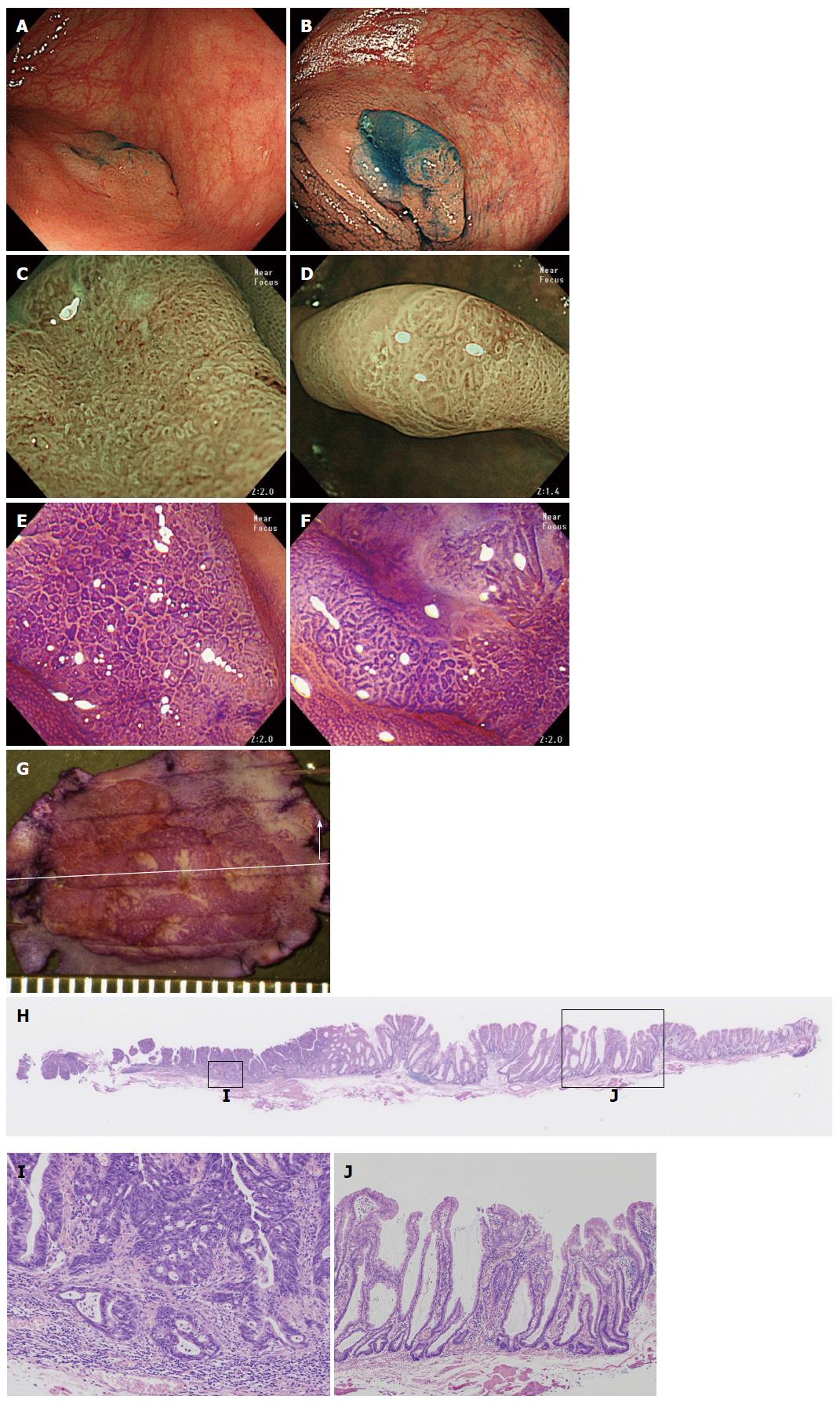
Figure 4 A case of an sessile serrated adenoma/polyp that has invaded the submucosal layer (scope: CF: HQ290I).
A: Conventional white light observation. A flat elevated polyp of approximately 20 mm with a reddish depressed area can be observed in the ascending colon; B: Indigocarmine spraying endoscopic finding. Chromoendoscopy revealed this lesion, which is clearly composed of lesions. One edge area is covered with thick mucus; C: Magnified NBI observation. Firmly attached mucus can be observed on the tumor. A II-D pit that is indicative are markedly dilated crypts can be seen in this area; D: Magnified NBI observation. A granular surface pattern with dilated microcapillary vessels can be observed on this tumor in the absence of a thick mucous adhesion; E and F: Magnified crystal violet staining observation; G: Stereoscopic finding. The tumor was excised by the EMR method. The tumor was cut into seven pieces; H: HE staining, whole specimen finding from #4; I: High power view of the HE staining. The neoplastic glands have invaded into the SM layer to a depth of approximately 400 μm. The glands exhibit high grade dysplastic change; J: Low power view of the HE staining. This polyp is composed of SSA/P glands with markedly dilated crypts. NBI: Narrow band imaging; SSA/P: Sessile serrated adenoma/polyp.
- Citation: Saito S, Tajiri H, Ikegami M. Serrated polyps of the colon and rectum: Endoscopic features including image enhanced endoscopy. World J Gastrointest Endosc 2015; 7(9): 860-871
- URL: https://www.wjgnet.com/1948-5190/full/v7/i9/860.htm
- DOI: https://dx.doi.org/10.4253/wjge.v7.i9.860


