Copyright
©The Author(s) 2015.
World J Gastrointest Endosc. Jul 25, 2015; 7(9): 860-871
Published online Jul 25, 2015. doi: 10.4253/wjge.v7.i9.860
Published online Jul 25, 2015. doi: 10.4253/wjge.v7.i9.860
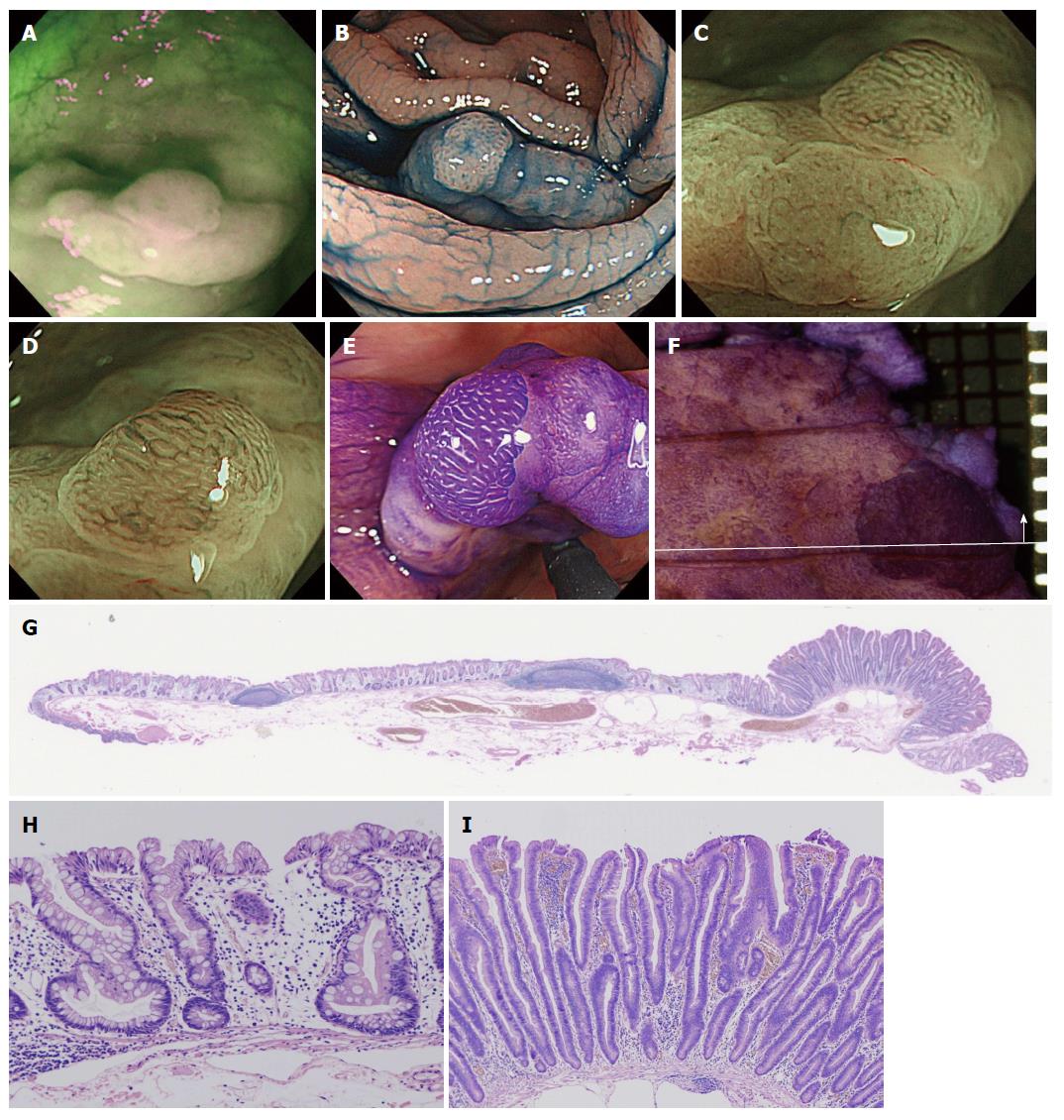
Figure 3 A case of sessile serrated adenoma/polyp with cytological dysplasia (scope: CF: FH260AZI).
A: AFI imaging. The polyp is shown as a flat elevated lesion with a small nodule and is located in the ascending colon. A slightly change to a magenta color can be seen localized to a small elevated lesion in the tumor; B: Indigocarmine spraying endoscopic finding. The small elevated nodule in the tumor can be seen observed following dye spraying; C: Magnified NBI observation. In the tumor lesion, whitish mucosa with II-D pits can be observed. The microcapillary vessels are not dilated in the tumor; D: Magnified NBI observation. In contrast, the microcapillary vessels are dilated surrounding the tumor pits at the small elevated nodule. Moreover, a IIIL pit (white line) can be indirectly observed; E: Magnified crystal violet staining observation. Type II open pits (II-O pits) containing normal type II pits are shown in the tumor; F: Stereoscopic finding. The tumor was excised by the ESD method. The tumor was cut eight pieces; G: HE staining, whole specimen findings from section #4 including a small nodule; H: High power view of the HE staining finding. A part of an SSA/P is shown in the picture; I: High power view of the HE staining finding. The small elevated lesion is shown as a neoplastic change. Low grade cytologic dysplasia is present with nuclear hyperchromasia and pseudostratification. NBI: Narrow band imaging; SSA/P: Sessile serrated adenoma/polyp; AFI: Auto fluorescence imaging.
- Citation: Saito S, Tajiri H, Ikegami M. Serrated polyps of the colon and rectum: Endoscopic features including image enhanced endoscopy. World J Gastrointest Endosc 2015; 7(9): 860-871
- URL: https://www.wjgnet.com/1948-5190/full/v7/i9/860.htm
- DOI: https://dx.doi.org/10.4253/wjge.v7.i9.860


