Copyright
©The Author(s) 2015.
World J Gastrointest Endosc. Jul 10, 2015; 7(8): 777-789
Published online Jul 10, 2015. doi: 10.4253/wjge.v7.i8.777
Published online Jul 10, 2015. doi: 10.4253/wjge.v7.i8.777
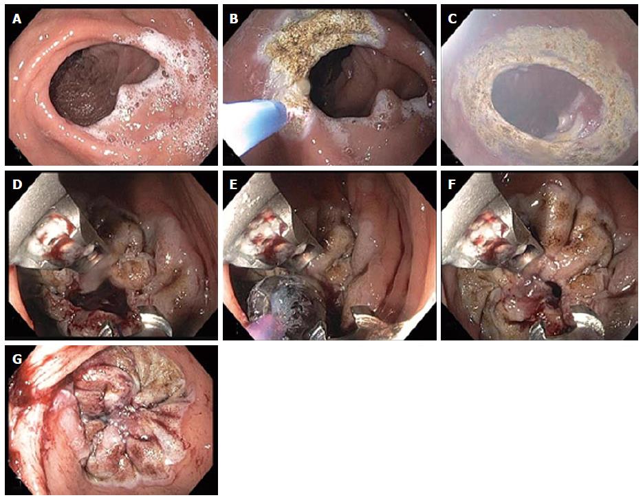
Figure 6 Endoscopic revision of gastrojejunal anastomosis in gastric bypass patient.
A: An enlarged gastrojejunal anastomosis is noted; B and C: Argon plasma coagulation was used around the stoma to ablate the mucosa and facilitate tissue fusion during the healing process; D: Two sutures were used obtaining circumferential tissue bites to achieve a purse-like closure of the stoma; E: A 10 mm controlled radial expansion balloon was dilated and placed through the stoma opening via the second channel of the double-channel therapeutic endoscope and then the sutures were tightened so that the final stoma diameter was approximately 10 mm in size; F and G: The balloon was then deflated and removed. A markedly diminished stoma orifice is seen at the end of the procedure.
- Citation: Stavropoulos SN, Modayil R, Friedel D. Current applications of endoscopic suturing. World J Gastrointest Endosc 2015; 7(8): 777-789
- URL: https://www.wjgnet.com/1948-5190/full/v7/i8/777.htm
- DOI: https://dx.doi.org/10.4253/wjge.v7.i8.777


