Copyright
©The Author(s) 2015.
World J Gastrointest Endosc. Jul 10, 2015; 7(8): 777-789
Published online Jul 10, 2015. doi: 10.4253/wjge.v7.i8.777
Published online Jul 10, 2015. doi: 10.4253/wjge.v7.i8.777
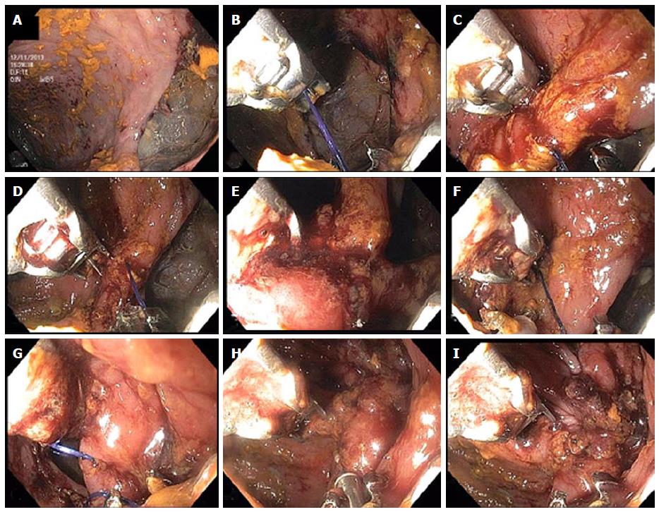
Figure 3 Closure of colonic perforation with endoscopic suturing device.
A: Initial tissue bites forming a running suture (B-E) starting at the inferior edge of the perforation and progressing towards the center; F: Tissue helix retractor is used to ensure deep tissue bite along the distal, superior edge of the perforation; G: Suturing has reached the superior edge of the perforation the edges of which are now being pulled together by the sutures; H: After tightening of the sutures closure of the perforation has been achieved and the cinch device is seen being deployed at the 6 o’ clock position of the image; I: Immediately after cinch deployment, the complete closure of the perforation is seen. Gastrograffin was injected through the scope that confirmed absence of leak (not shown).
- Citation: Stavropoulos SN, Modayil R, Friedel D. Current applications of endoscopic suturing. World J Gastrointest Endosc 2015; 7(8): 777-789
- URL: https://www.wjgnet.com/1948-5190/full/v7/i8/777.htm
- DOI: https://dx.doi.org/10.4253/wjge.v7.i8.777


