Copyright
©The Author(s) 2015.
World J Gastrointest Endosc. Jul 10, 2015; 7(8): 758-768
Published online Jul 10, 2015. doi: 10.4253/wjge.v7.i8.758
Published online Jul 10, 2015. doi: 10.4253/wjge.v7.i8.758
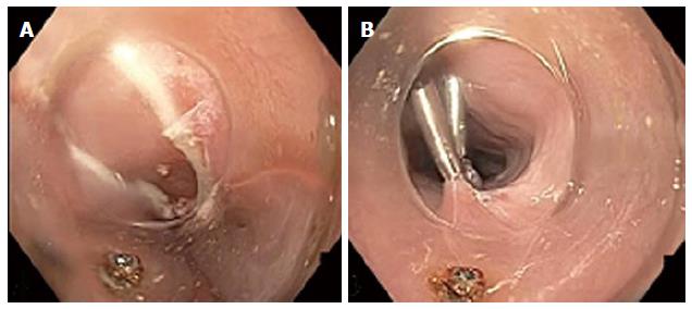
Figure 2 Endoscopic view of mucosotomy during peroral endoscopic myotomy.
A: Esophageal mucosal defect after completion of peroral endoscopic myotomy; B: Defect closed with sequentially placed through the scope clips.
- Citation: Winder JS, Pauli EM. Comprehensive management of full-thickness luminal defects: The next frontier of gastrointestinal endoscopy. World J Gastrointest Endosc 2015; 7(8): 758-768
- URL: https://www.wjgnet.com/1948-5190/full/v7/i8/758.htm
- DOI: https://dx.doi.org/10.4253/wjge.v7.i8.758


