Copyright
©The Author(s) 2015.
World J Gastrointest Endosc. Jun 25, 2015; 7(7): 688-701
Published online Jun 25, 2015. doi: 10.4253/wjge.v7.i7.688
Published online Jun 25, 2015. doi: 10.4253/wjge.v7.i7.688
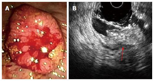
Figure 3 Stage T3 rectal cancer: (A) endoscopic and (B) ultrasonographic view.
Endoscopic ultrasound with radial array transducer UM160 (5-20 MHz), showing increased wall thickness for the presence of a mass with inhomogeneous echogenicity, invading all the layers of the wall and minimal infiltration of the perirectal fat. T: Tumor; Red arrow: Infiltration of the perirectal fat.
- Citation: Marone P, Bellis M, D’Angelo V, Delrio P, Passananti V, Girolamo ED, Rossi GB, Rega D, Tracey MC, Tempesta AM. Role of endoscopic ultrasonography in the loco-regional staging of patients with rectal cancer. World J Gastrointest Endosc 2015; 7(7): 688-701
- URL: https://www.wjgnet.com/1948-5190/full/v7/i7/688.htm
- DOI: https://dx.doi.org/10.4253/wjge.v7.i7.688


