Copyright
©The Author(s) 2015.
World J Gastrointest Endosc. Jun 10, 2015; 7(6): 575-581
Published online Jun 10, 2015. doi: 10.4253/wjge.v7.i6.575
Published online Jun 10, 2015. doi: 10.4253/wjge.v7.i6.575
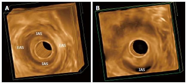
Figure 2 Three-dimensional endoanal ultrasonography images.
A: Normal appearance of the external anal sphincter (EAS) and internal anal sphincter (IAS); B: An IAS defect in woman as a complication of a previous anorectal surgery (due to fistula).
- Citation: Albuquerque A. Endoanal ultrasonography in fecal incontinence: Current and future perspectives. World J Gastrointest Endosc 2015; 7(6): 575-581
- URL: https://www.wjgnet.com/1948-5190/full/v7/i6/575.htm
- DOI: https://dx.doi.org/10.4253/wjge.v7.i6.575


