Copyright
©The Author(s) 2015.
World J Gastrointest Endosc. May 16, 2015; 7(5): 567-572
Published online May 16, 2015. doi: 10.4253/wjge.v7.i5.567
Published online May 16, 2015. doi: 10.4253/wjge.v7.i5.567
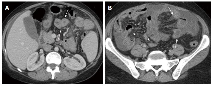
Figure 4 Triphasic post contrast axial computed tomography (portal phase) showing.
A: Dilated small intestinal wall (arrows); B: Mesenteric hypodense bands indicating obstructed lymphatics (arrows), and dirty fat appearance due to mesenteric oedema (arrow heads).
- Citation: El-Etreby SA, Altonbary AY, Sorogy ME, Elkashef W, Mazroa JA, Bahgat MH. Anaemia in Waldmann’s disease: A rare presentation of a rare disease. World J Gastrointest Endosc 2015; 7(5): 567-572
- URL: https://www.wjgnet.com/1948-5190/full/v7/i5/567.htm
- DOI: https://dx.doi.org/10.4253/wjge.v7.i5.567


