Copyright
©The Author(s) 2015.
World J Gastrointest Endosc. May 16, 2015; 7(5): 563-566
Published online May 16, 2015. doi: 10.4253/wjge.v7.i5.563
Published online May 16, 2015. doi: 10.4253/wjge.v7.i5.563
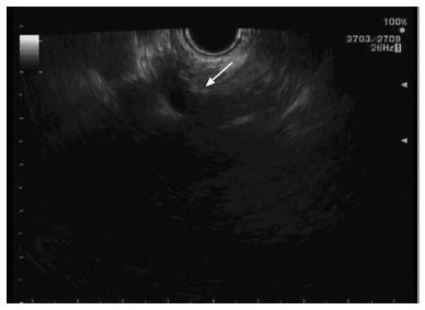
Figure 3 Ultrasonographic image of celiac plexus.
This image is showing the celiac artery and celiac plexus (arrow).
- Citation: Alhazzani W, Al-Shamsi HO, Greenwald E, Radhi J, Tse F. Chronic abdominal pain secondary to mesenteric panniculitis treated successfully with endoscopic ultrasonography-guided celiac plexus block: A case report. World J Gastrointest Endosc 2015; 7(5): 563-566
- URL: https://www.wjgnet.com/1948-5190/full/v7/i5/563.htm
- DOI: https://dx.doi.org/10.4253/wjge.v7.i5.563


