Copyright
©The Author(s) 2015.
World J Gastrointest Endosc. Apr 16, 2015; 7(4): 396-402
Published online Apr 16, 2015. doi: 10.4253/wjge.v7.i4.396
Published online Apr 16, 2015. doi: 10.4253/wjge.v7.i4.396
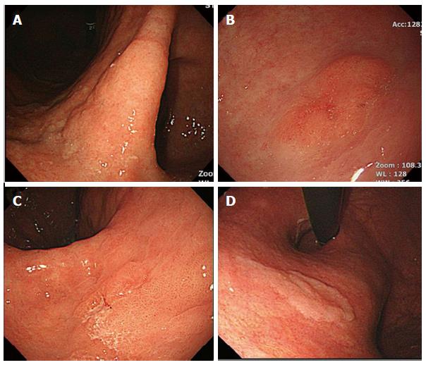
Figure 2 Endoscopic images of biopsy-proven low-grade dysplasia.
A-C: lesion size > 2 cm (A), surface erythema (B), and depressed appearance (C) are endoscopic risk factors for an upgraded histology after endoscopic resection; D: In contrast, the presence of whitish discoloration was a negative factor.
- Citation: Kim JW, Jang JY. Optimal management of biopsy-proven low-grade gastric dysplasia. World J Gastrointest Endosc 2015; 7(4): 396-402
- URL: https://www.wjgnet.com/1948-5190/full/v7/i4/396.htm
- DOI: https://dx.doi.org/10.4253/wjge.v7.i4.396


