Copyright
©The Author(s) 2015.
World J Gastrointest Endosc. Apr 16, 2015; 7(4): 318-327
Published online Apr 16, 2015. doi: 10.4253/wjge.v7.i4.318
Published online Apr 16, 2015. doi: 10.4253/wjge.v7.i4.318
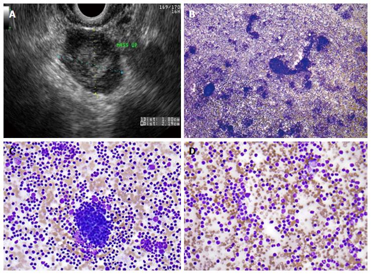
Figure 3 Primary pancreatic lymphoma.
A: Endoscopic ultrasound demonstrating a 1.8 cm × 2.2 cm lymphoma in the uncinate process of the pancreas; B: Low-power view showing a very cellular aspirate composed of discohesive lymphoid cells (Diff-QuikTM stain, × 100); C: High-power view showing an admixture of mature lymphocytes of various sizes with no more than a minimal atypia; lymphoid aggregates resembling a germinal center are also present (bottom); D: Small mature lymphocytes with cleaved and irregular nuclei raising suspicion for a mature B-cell lymhoma. (Diff-QuikTM stain, × 400).
- Citation: Nelsen EM, Buehler D, Soni AV, Gopal DV. Endoscopic ultrasound in the evaluation of pancreatic neoplasms-solid and cystic: A review. World J Gastrointest Endosc 2015; 7(4): 318-327
- URL: https://www.wjgnet.com/1948-5190/full/v7/i4/318.htm
- DOI: https://dx.doi.org/10.4253/wjge.v7.i4.318


