Copyright
©The Author(s) 2015.
World J Gastrointest Endosc. Apr 16, 2015; 7(4): 318-327
Published online Apr 16, 2015. doi: 10.4253/wjge.v7.i4.318
Published online Apr 16, 2015. doi: 10.4253/wjge.v7.i4.318
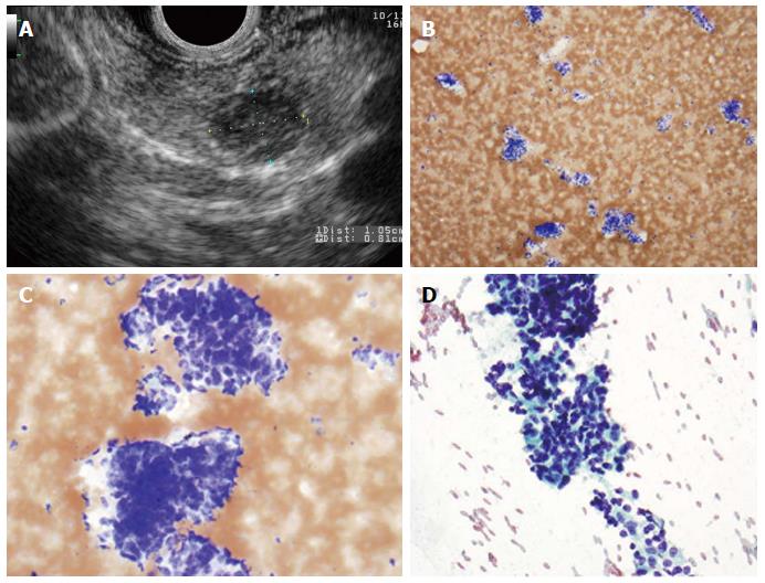
Figure 2 Pancreatic neuroendocrine neoplasm.
A: Endoscopic ultrasound image showing a 9 mm × 10 mm neuroendocrine tumor (insulinoma); B: Low-power view shows a cellular aspirate composed of clusters of uniform cells (Diff-QuikTM stain, × 100); C: High power view shows uniform cells with high N:C ratios and coarse chromatin (Diff-QuikTM stain, × 400); D: Papanicolaou stain highlights coarse, evenly distributed chromatin (× 400).
- Citation: Nelsen EM, Buehler D, Soni AV, Gopal DV. Endoscopic ultrasound in the evaluation of pancreatic neoplasms-solid and cystic: A review. World J Gastrointest Endosc 2015; 7(4): 318-327
- URL: https://www.wjgnet.com/1948-5190/full/v7/i4/318.htm
- DOI: https://dx.doi.org/10.4253/wjge.v7.i4.318


