Copyright
©The Author(s) 2015.
World J Gastrointest Endosc. Mar 16, 2015; 7(3): 224-229
Published online Mar 16, 2015. doi: 10.4253/wjge.v7.i3.224
Published online Mar 16, 2015. doi: 10.4253/wjge.v7.i3.224
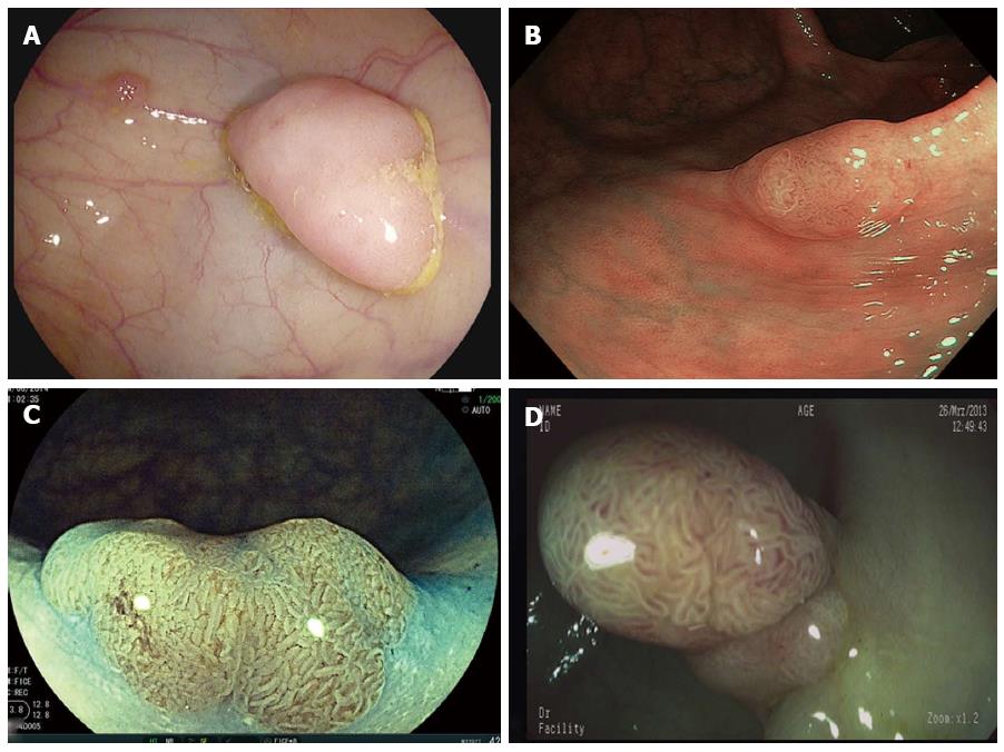
Figure 1 Colonic polyp imaged with high-definition white-light (A), narrow band imaging (B), Fujinon Intelligent Color Enhancement (C) and i-scan (D).
Data on detection rates are inconsistent. Nevertheless, dye-less chromoendoscopy techniques allow for a detailed and adequate examination of the mucosal pit pattern and the mucosal vascular pattern morphology to predict polyp histology in real time.
- Citation: Neumann H, Nägel A, Buda A. Advanced endoscopic imaging to improve adenoma detection. World J Gastrointest Endosc 2015; 7(3): 224-229
- URL: https://www.wjgnet.com/1948-5190/full/v7/i3/224.htm
- DOI: https://dx.doi.org/10.4253/wjge.v7.i3.224


