Copyright
©The Author(s) 2015.
World J Gastrointest Endosc. Mar 16, 2015; 7(3): 192-205
Published online Mar 16, 2015. doi: 10.4253/wjge.v7.i3.192
Published online Mar 16, 2015. doi: 10.4253/wjge.v7.i3.192
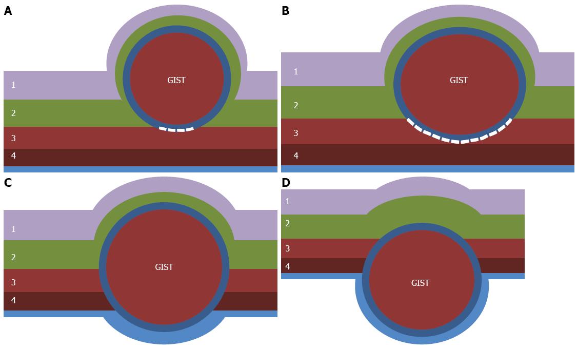
Figure 2 Classification of gastrointestinal stromal tumors according to the location in the gastric wall.
A: Type I is a gastrointestinal stromal tumor (GIST) that has a very narrow connection with the proper muscle layer and protrudes into the luminal side like a polyp; B: Type II has a wider connection with the proper muscle layer and protrudes into the luminal side at an obtuse angle; C: Type III is located in the middle of the gastric wall; D: Type IV protrudes mainly into the serosal side of the gastric wall. White dotted lines indicate the area dissected from the proper muscle layer. 1: Mucosa; 2: Submucosa; 3: Circular layer of proper muscle; 4: Longitudinal layer of proper muscle.
- Citation: Kim HH. Endoscopic treatment for gastrointestinal stromal tumor: Advantages and hurdles. World J Gastrointest Endosc 2015; 7(3): 192-205
- URL: https://www.wjgnet.com/1948-5190/full/v7/i3/192.htm
- DOI: https://dx.doi.org/10.4253/wjge.v7.i3.192


