Copyright
©The Author(s) 2015.
World J Gastrointest Endosc. Feb 16, 2015; 7(2): 110-120
Published online Feb 16, 2015. doi: 10.4253/wjge.v7.i2.110
Published online Feb 16, 2015. doi: 10.4253/wjge.v7.i2.110
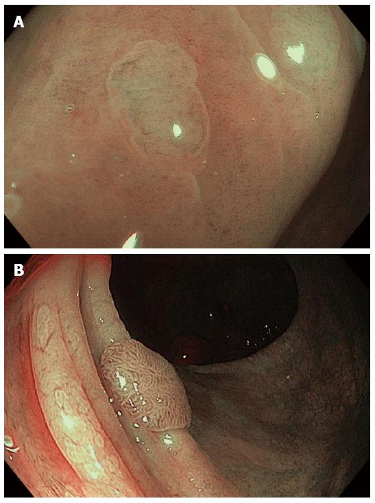
Figure 7 Assessment of colonic lesions by narrow band imaging with magnification endoscopy according to different classification systems.
A: Hyperplastic polyp: absent mesh brown capillary network (Type I MBCN) (Sano classification); a lighter color of the polyp than the background, isolated vessels coursing across the lesion (NICE criteria); B: Adenomatous polyp: regular mesh brown capillary network (Type II MBCN) and Kudo’s Type IV mucosal pattern; the brown color relative to background, thick brown vessels surrounding white structures (NICE criteria); C: Cancerous colonic lesion: irregular mucosal and vascular patterns (Type III MBCN); D: Deep submucosal invasive colorectal cancer: amorphous surface pattern and disrupted vessels (NICE criteria).
- Citation: Boeriu A, Boeriu C, Drasovean S, Pascarenco O, Mocan S, Stoian M, Dobru D. Narrow-band imaging with magnifying endoscopy for the evaluation of gastrointestinal lesions. World J Gastrointest Endosc 2015; 7(2): 110-120
- URL: https://www.wjgnet.com/1948-5190/full/v7/i2/110.htm
- DOI: https://dx.doi.org/10.4253/wjge.v7.i2.110


