Copyright
©The Author(s) 2015.
World J Gastrointest Endosc. Feb 16, 2015; 7(2): 110-120
Published online Feb 16, 2015. doi: 10.4253/wjge.v7.i2.110
Published online Feb 16, 2015. doi: 10.4253/wjge.v7.i2.110
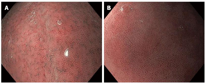
Figure 2 Narrow band imaging with magnification endoscopy images of normal gastric mucosa.
A: Round pits surrounded by the subepithelial capillary network (SECN) and collecting venules (CVs) in normal corporeal mucosa; B: Coil-shaped appearance of SECN, without the visualization of the CVs in normal antral mucosa.
- Citation: Boeriu A, Boeriu C, Drasovean S, Pascarenco O, Mocan S, Stoian M, Dobru D. Narrow-band imaging with magnifying endoscopy for the evaluation of gastrointestinal lesions. World J Gastrointest Endosc 2015; 7(2): 110-120
- URL: https://www.wjgnet.com/1948-5190/full/v7/i2/110.htm
- DOI: https://dx.doi.org/10.4253/wjge.v7.i2.110


