Copyright
©The Author(s) 2015.
World J Gastrointest Endosc. Feb 16, 2015; 7(2): 110-120
Published online Feb 16, 2015. doi: 10.4253/wjge.v7.i2.110
Published online Feb 16, 2015. doi: 10.4253/wjge.v7.i2.110
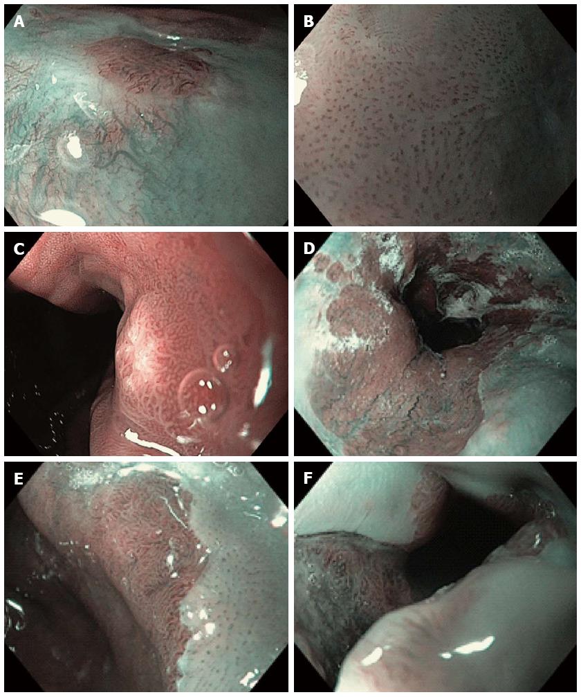
Figure 1 Narrow band imaging with magnification endoscopy images of the esophagus.
A: Normal esophageal mucosa: branching vessel network and intra-epithelial papillary capillary loop (IPCL) surrounding an island of Barrett’s esophagus (BE); B: IPCL type V1: dilatation of intra-epithelial papillary capillary loop, irregular caliber, and form variation; C: Round pits with regular microvasculature corresponding with columnar mucosa; D: Non-dysplastic BE: flat-type mucosa with regular long branching vessels; E: Non-dysplastic BE: regular villous/ridge mucosal pattern; F: Dysplastic BE: distorsion of mucosal pattern and irregular vascular pattern.
- Citation: Boeriu A, Boeriu C, Drasovean S, Pascarenco O, Mocan S, Stoian M, Dobru D. Narrow-band imaging with magnifying endoscopy for the evaluation of gastrointestinal lesions. World J Gastrointest Endosc 2015; 7(2): 110-120
- URL: https://www.wjgnet.com/1948-5190/full/v7/i2/110.htm
- DOI: https://dx.doi.org/10.4253/wjge.v7.i2.110


